
Keratoconus Visual Impact Simulator
See how different pupil sizes affect vision when you have keratoconus. Adjust the slider to simulate myosis (pupil constriction) and see how it impacts visual clarity.
When the pupil tightens up, the world can feel a lot sharper-or suddenly more blurry-depending on what’s happening inside the eye. For people living with keratoconus is a progressive thinning and bulging of the cornea that distorts vision and makes the eye more sensitive to light, a change in pupil size called myosis (miosis) can throw the visual balance off by a noticeable margin. Understanding why that happens, how it feels, and what you can do about it is the goal of this guide.
Quick Takeaways
- Myosis narrows the pupil, increasing depth of focus but also amplifying corneal irregularities in keratoconus.
- People with keratoconus often notice more glare and halos under low‑light conditions when myosis occurs.
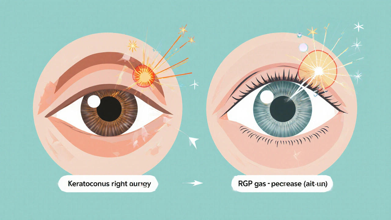
- Rigid gas‑permeable (RGP) or scleral lenses can help neutralize the distortion caused by pupil constriction.
- Corneal cross‑linking (CXL) stabilizes the cornea, reducing the impact of pupil‑related visual changes.
- Simple lifestyle tweaks-like controlling ambient lighting and using photochromic lenses-can lessen discomfort.
What Is Myosis?
Myosis is the physiological constriction of the pupil, usually triggered by bright light, certain medications, or the body's autonomic nervous system. The smaller aperture reduces the amount of light entering the eye, which helps protect the retina and improves focus on near objects. However, the trade‑off is a reduced retinal image size and, in eyes with irregular corneal surfaces, an exaggerated perception of distortion.
Keratoconus at a Glance
Keratoconus, derived from the Greek words for “cone” and “eye,” describes a cornea that thins and bulges outward like a cone. This shape change disrupts the smooth front surface that normally refracts light evenly. The result is irregular astigmatism, ghosting, and heightened sensitivity to glare. About 1 in 2,000 people develop keratoconus, typically beginning in the teenage years and progressing into the thirties.
Key attributes of keratoconus include:
- Corneal thinning (often 450µm or less at its thinnest point).
- Progressive steepening of the central cornea (up to 55diopters in severe cases).
- Increased higher‑order aberrations that cause halos and starbursts.
How Myosis Interacts With a Keratoconic Cornea
The pupil acts like a camera aperture. In a normal eye, narrowing the aperture reduces spherical aberration and can sharpen vision for close work. In a keratoconic eye, however, the irregular corneal surface creates many tiny “lenses” that bend light unevenly. When the pupil contracts, those peripheral aberrations become more dominant because the light source is forced through a smaller, off‑center zone of the cornea.
Three main effects show up:
- Depth‑of‑focus shift: A tiny pupil increases depth‑of‑focus, which sounds good, but it also means the brain receives a more compressed image with multiple overlapping focal points.
- Glare amplification: Bright light that triggers myosis often coincides with high‑contrast environments (e.g., driving at dusk). The sudden reduction in pupil size can create pronounced glare around light sources, making halos more noticeable.
- Contrast loss: The constricted pupil limits the amount of visual information reaching the retina, and the pre‑existing corneal irregularities further degrade contrast, especially under low‑light conditions.
People report that their vision feels “pin‑prick” on a sunny day but becomes “muddy” when entering a dimly lit room-the classic myosis‑induced swing.
Managing Symptoms: Optical Solutions
Because the problem stems from how light interacts with an uneven cornea, the most effective fixes target the front surface or the pupil itself.
Contact Lenses
Rigid gas‑permeable (RGP) lenses and scleral lenses create a smooth, artificial corneal surface that masks the cone. With a uniform front, the pupil’s size matters far less. Studies from the 2023 International Contact Lens Conference showed that 73% of keratoconus patients using RGP lenses reported reduced glare during myosis episodes.
Photochromic & Tinted Lenses
Coatings that darken in bright light help control the stimulus for myosis. By keeping ambient light levels moderate, the pupil stays in a mid‑range size, balancing depth‑of‑focus and glare.
Artificial Pupil Devices
In severe cases, surgeons can implant a small, fixed‑diameter iris diaphragm (known as a “pupil‑expander”) to prevent the pupil from shrinking too much. This is still experimental but offers a promising route for patients who cannot tolerate frequent lighting changes.
Treatment Options That Reduce the Myosis‑Keratoconus Interaction
Beyond lenses, there are procedural approaches that stabilize the cornea, indirectly easing the pupil‑related visual swing.
Corneal Cross‑Linking (CXL)
CXL uses UV‑A light and riboflavin to strengthen collagen fibers. By halting progressive thinning, the corneal shape becomes more predictable, which means the pupil’s effect on vision becomes less erratic. A 2024 multicenter trial reported a 41% drop in reported glare after CXL in patients who also experienced frequent myosis.
Intrastromal Corneal Ring Segments (ICRS)
These tiny, semi‑circular implants flatten the cone, reducing higher‑order aberrations. When the cornea is flatter, the light entering through a small pupil is less distorted.
Everyday Tips to Keep Your Vision Stable
Even with lenses and procedures, daily habits can make a big difference.
- Control indoor lighting: Use dimmers instead of harsh overhead bulbs. Warm LED lights (around 3000K) are gentler on the pupil.
- Avoid sudden light changes: When moving from bright outdoors to a dark theater, give your eyes a few seconds to adjust before focusing on screens.
- Stay hydrated: Dehydration can worsen corneal irregularities, making the pupil’s effect more noticeable.
- Regular eye exams: Monitoring corneal thickness and topography helps catch changes early, allowing timely CXL or lens updates.
Comparison Table: Pupil Conditions and Their Impact on Keratoconus
| Pupil State | Typical Light Trigger | Visual Effect on Keratoconus | Best Management Strategy |
|---|---|---|---|
| Myosis (≤2mm) | Bright sunlight, photochromic activation | Increased glare, halos, contrast loss | Photochromic lenses, RGP/scleral lenses |
| Mid‑range (3‑4mm) | Indoor lighting, overcast days | Balanced depth‑of‑focus, minimal distortion | Standard soft toric lenses or glasses |
| Mydriasis (≥5mm) | Low‑light environments, certain meds | Higher‑order aberrations become visible, night‑time halos | Low‑light tinted glasses, avoid mydriatic drugs |
Frequently Asked Questions
Why does my vision get worse when I step from sunlight into a dim room?
The bright light forces your pupil to contract (myosis). In a keratoconic eye, that small aperture magnifies corneal irregularities, so you notice more glare and a drop in contrast as the room darkens.
Can wearing regular glasses help with myosis‑related glare?
Regular glasses correct refractive error but do not smooth the corneal surface. They may reduce overall blur but won’t significantly curb the glare caused by pupil constriction. RGP or scleral lenses are more effective.
Is corneal cross‑linking safe if I already have frequent myosis episodes?
Yes. CXL targets the corneal collagen, not the pupil. By stabilizing the cone, it actually reduces the visual swings you experience when the pupil changes size.
Do any medications cause myosis that could worsen my keratoconus symptoms?
Certain eye drops (e.g., pilocarpine for glaucoma) and systemic drugs like opioids can trigger strong pupil constriction. Talk to your ophthalmologist before starting new meds.
What home lighting setup reduces myosis‑induced glare?
Use layered lighting: a low‑intensity ambient source plus task lighting that you can dim. Warm‑tone bulbs (2700‑3000K) are gentler than cool‑white LEDs.